I hope you found my previous article on BN-PAGE useful. In this post, I will continue the focus on gel electrophoresis and discuss the various methods for western blot detection. This includes how they work as well as the advantages and disadvantages of each. Let's begin!
What Are the Existing Detection Methods for Western Blot?
The main techniques for visualizing a western blot are colorimetric, chemiluminescence, and fluorescence.
Colorimetric and chemiluminescence act by an enzymatic reaction either by horseradish peroxidase or alkaline phosphatase (also used in ELISA). Meanwhile, fluorescence methods do not depend on a reaction but instead rely on fluorophore-conjugated antibodies that can be visualized directly.
Detection of your target protein typically involves two antibodies, a primary antibody against your target protein and a secondary antibody against the host species of your primary that’s been conjugated to an enzyme or a fluorophore.
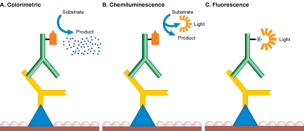
Image Credit: Bio-Rad
Colorimetric
Advantages: Cheap, easy to use, quick results.
Disadvantages: Low sensitivity (picogram range), only single output signal.
Colorimetric assays act by producing a colored precipitate from an enzymatic reaction. For example, HRP catalyzes a reaction with 4-Chloro-1-napthol (4CN) and peroxide that produces a visible and insoluble purple product. Colorimetric assays are generally easier to use and don’t require the costly materials of X-ray film or a developer. However, their range of sensitivity requires 5-500 pg of protein; much higher than chemiluminescent sensitivity, which can be in the femtogram range.
I personally have never witnessed any current research labs that still use colorimetric detection methods for western blotting (I’m sure there’s probably a few out there still). Though I believe it may still be used for undergraduate laboratory classes. Given the low sensitivity of colorimetric methods, I would only recommend these techniques for non-quantitative purposes with highly expressed proteins.
Chemiluminescence
Detection method: Film
Advantages: High sensitivity (femtogram range).
Disadvantages: Non-linear, small range of detection, single output signal, requires dark room, continuous cost of film and developer maintenance.
Detection method: CCD
Advantages: Easy to use and more linear than film.
Disadvantages: Possibly less sensitive than film, cost of a CCD, single output signal.
Chemiluminescence works by producing light as a byproduct from a reaction between a chemical substrate and the enzyme. Light intensity can then be measured by exposure to an X-ray film or a charged coupled device (CCD) that converts photons to an electronic signal. Between the enzymatic methods of western blot detection, chemiluminescence is far more sensitive and more commonly used than colorimetric methods. In fact, chemiluminescence coupled with X-ray film is the most sensitive method for western blot detection. Given the low expression rates of some of the studied proteins in our lab, we have yet to see comparable sensitivity with a CCD under the same blotting conditions and exposure time.
This being said, X-ray film lacks the linearity and range of detection as a CCD. For these reasons, X-ray film should never be used for quantitative purposes and a normalized loading control should always be present (i.e. a housekeeping gene or protein that should not vary between samples given the treatment, such as GAPDH or beta-Actin). Furthermore, given the limited detection range of X-ray film, an underexposed film should always be used to ensure proper comparison of band intensity between lanes. I will discuss technique and proper troubleshooting further in part 2 of this series.
Fluorescence
Advantages: Wide range of detection, linear, quantifiable, easy to use, and multiple output signals with different fluorophores.
Disadvantages: May not be as sensitive as chemiluminescence and film, cost of an imager.
Lastly, fluorescence is the more widely used and versatile Western detection method these days. Instead of an enzymatic reaction producing a signal, antibodies are conjugated to a specific fluorophore and can be detected using an imaging system. For this reason, fluorescence detection methods have a far greater range of linearity than using film with chemiluminescence. Fluorescence also has the advantage of simultaneously probing multiple proteins with various fluorophores. This is a huge advantage over chemiluminescent methods that require a stripping and re-probing protocol for proteins that run at a similar molecular weight.
Stripping a blot requires a second round of imaging and can reduce the signal from the membrane. Several imaging systems are available for fluorescent Western blotting that excites fluorophores either by visible light, LED, or infrared lasers.
Odyssey systems from Li-Cor are a generally favored brand and offer infrared laser excitation which has the most narrow excitation window for the greatest reduction in background noise.
For additional information on these detection methods, check out this western blot guide by Science.
In my next article, I will elaborate on tips for using X-ray film for chemiluminescence western blot. Until then!
If you're planning a western blot experiment, try BenchSci and filter for published western blot data to help with your antibody search.