
CAR-T (chimeric antigen receptor (CAR)-engineered T cells) therapy is a new and promising anti-cancer immunotherapy modality. Among CAR-T cell therapies, CD19 CAR-T therapy, which involves genetically engineering autologous T cells ex vivo to express CARs against a B-lineage antigen CD19, seems arguably the most successful. Results obtained with CD19 CAR-T cell therapy in patients with B-cell tumors, including diffuse large B-cell lymphoma (DLBCL) and B-cell precursor acute lymphocytic leukemia (B-ALL), showed promising but variable efficacy.
To identify the key contributors to these variations, a recent study by Xue et al. proposed a proteomic approach based on a single-cell, 16-plex cytokine microfluidic device to characterize the functional attributes of CD19 CAR-T pre-infusion products ex vivo. Their method analysis is based on the finding that polyfunctional T cells co-producing multiple cytokines/chemokines at the single cell level, as demonstrated by flow cytometry intracellular cytokine staining, were the key effector cells contributing to the development of potent and durable cellular immunity against viral infection or cancer. The study authors applied the same rationale but using a single-cell 16-plex cytokine microfluidic device, a proteomic method, for a higher resolution single-cell analysis of CD19 CAR-T pre-infusion products.
In this article, I will summarize the protocol of the novel method proposed by Xue et al. for your reference.
What Is a Single-cell 16-Plex Cytokine Microfluidic Device?
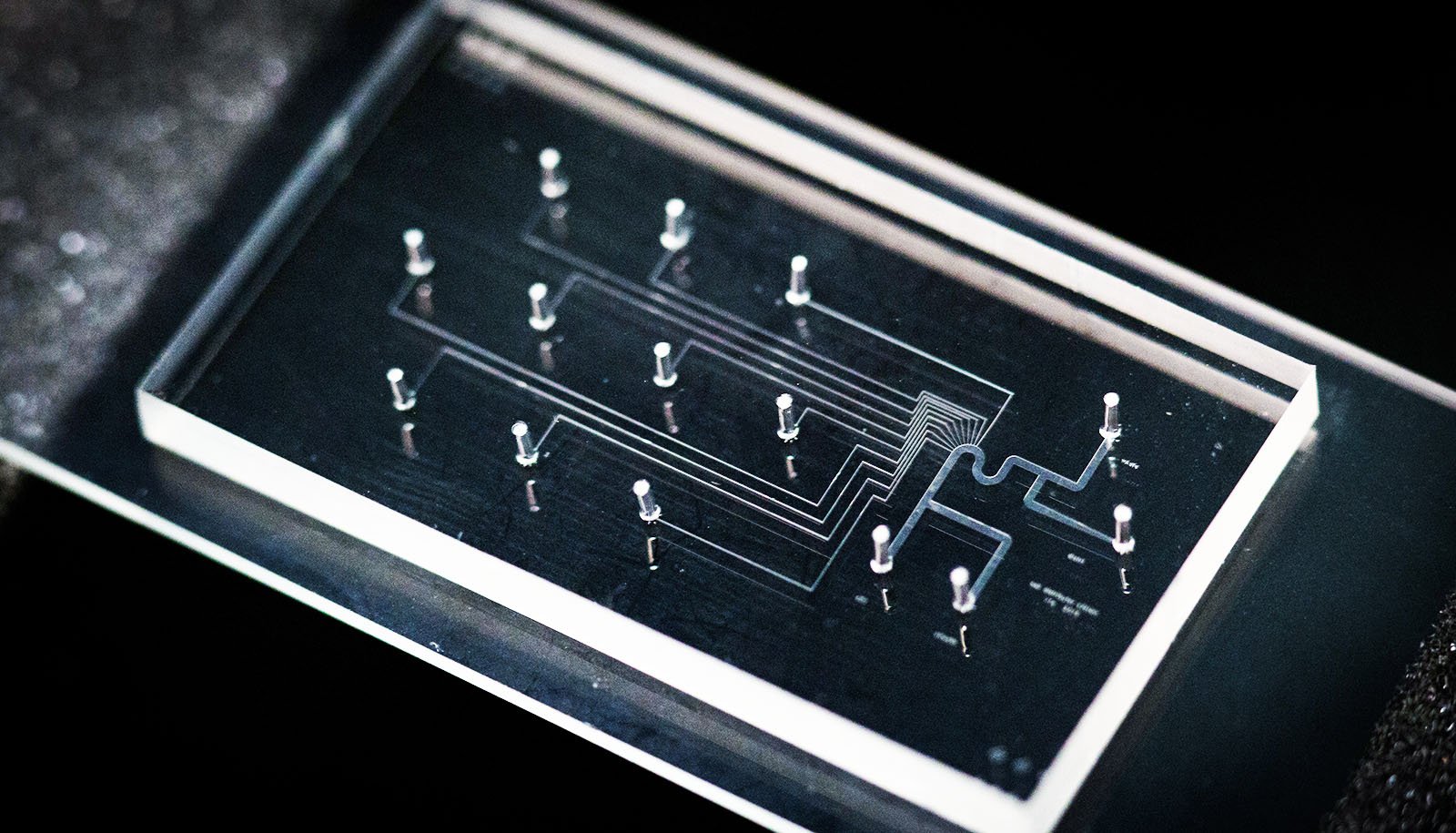
A single-cell 16-plex cytokine microfluidic device can be counted among newly emerging, highly multiplexed, single-cell microfluidic proteomic technologies. The device, which is a single-cell barcode chip microdevice, is made of multiple microchambers, covered with a glass slide pre-patterned with a complete copy of antibody array designed for capturing cytokine secretions from CAR-T cells following stimulations and staining for cell surface markers (e.g. CD4 and CD8) before being analyzed per cell type with the use of proprietary software (IsoPlexis’ IsoSpeak software package) and new visualization methods. The device in its standard format can simultaneously measure up to 42 cytokines at the single cell level. To conduct their study, Xue et al. used a 16-plex cytokine device format.
How Was the Study Conducted?
The study was carried out by the authors with the use of PBMC samples from the apheresis of 4 different healthy donors (2 females and 2 males with age range 26-51). The donor PBMC samples were transduced with a lentivirus encoding the CD19-BB-z transgene to generate CD19 CAR-T cells which were expanded using anti-CD3/anti-CD28 coated beads before being specifically stimulated with the antigen of interest, and stained for CD4 and CD8 markers and tested for cytokine secretions using a single-cell, 16-plex cytokine microfluidics device. Before initiating assays, the study authors firstly validated the microdevice at both the population and single-cell levels.
Important Protocol Features
To perform their experiments with the single-cell 16-plex cytokine microdevice, the authors applied this protocol.
- CAR-T cells were prepared by transducing with a lentivirus encoding the CD19-BB-z transgene healthy PBMCs isolated from the apheresis of healthy donors in a modified X-VIVO 15 media.
- The transfected cells were expanded with anti-CD3/anti-CD28 coated beads (at a 1:4 ratio) in a closed system for 10 days. Cell concentration was monitored during the expansion process and fresh medium was added every 2-3 days. On the day of harvest, transduced CAR-T cells were isolated from the population using Miltenyi anti-PE microbeads following the manufacture’s instructions.
- The enriched CAR-T cells (1 × 10⁶ cells/mL) were stimulated with anti-CAR or control IgG beads mixed (at a 1:4 ratio) in wells of a 96 well tissue culture plate at 37 °C for 6 h and then stained with anti-human CD4 RPE antibody (1:100 dilution) and antihuman CD8a Alexa Fluor 647 antibody (1:100 dilution) at room temperature for 10 min. The cells were then spun at 300 g for 10 min and re-suspended in fresh media at a density of 1.25 × 10⁶ cells/mL
- Following stimulation and staining, 30 μL of CAR-T cell suspension were then incubated for 16 h in a single-cell 16-plex cytokine microfluidic device. Protein secretions from single CAR-T cells were captured and subsequently analyzed using proprietary software and new visualization methods. The software identified and counted cells in each microchamber based on fluorescence intensities in the fluorescence images and feature detection in the bright field image. Cell subsets (CD4/CD8) were further determined based on immunofluorescence.
- To assess polyfunctional heterogeneity of activated CD19 CAR-T cells following the assay, the percentage of polyfunctional cells was computed at first regardless of the combination of cytokines co-produced and then per cytokine type using a polyfunctional strength index (PSI). Cytokines types including Effector (Granzyme B, IFN-G, MIP-1a, TNF-a), stimulatory (GM-CSF, IL-2, IL-8), regulatory (IL-4, IL-13, IL-22), and inflammatory (IL-6, IL-17) cytokines were monitored. PSI express the percentage of polyfunctional cells multiplied by the average signal intensity of the cytokines secreted by cells.
Study Findings and Conclusion
Following procedures as summarized in this article, the study authors found that:
- Less than 5% of cells with IgG bead stimulation exhibited polyfunctionality. By contrast, anti-CAR bead-stimulated CAR-T cells of the 4 donors showed 6-, 2-, 47-, and 11-fold increase in polyfunctional cell counts.
- Anti-CAR bead-stimulated CD8+ T cells seemed more polyfunctional than their CD4+ T cell counterparts.
- PSI values showed more contribution of effector and stimulatory cytokines to polyfunctionality across all 4 donors with a small portion of regulatory and inflammatory cytokines contributing in donor 1 and donor 4.
- Furthermore, the observed regulatory and inflammatory responses were mainly from the CD4+ T cells, consistent with the notion that both regulatory and helper T cells are subsets of CD4+ T cells, further proving the specificity of the assay.
Considering PSI results, the authors of this study concluded that CD19 CAR-T pre-infusion products from the 4 donors displayed a diversified landscape of immune effector response following antigen-specific challenge. The novel single-cell, 16-plex analysis system described by Xue et al provides a mean for a more sensitive and comprehensive functional assessment of CAR-T pre-infusion products, and may provide insights into the safety and efficacy of CAR-T cell therapies.
Editor's note: If you are studying any of the biomarkers mentioned in this article, we put together a list of antibodies with the most published figures on BenchSci for you to review.
| Target | Clonality | Host | View Published Data |
| CD3 | Polyclonal | Rabbit |  |
| CD4 | Monoclonal | Mouse |  |
| CD8a | Monoclonal | Rat |  |
| CD8b | Monoclonal | Mouse |  |
| CD19 | Monoclonal | Mouse |  |
| CD28 | Monoclonal | Mouse |  |
| IL-2 | Monoclonal | Rat |  |
| IL-4 | Monoclonal | Rat |  |
| IL-6 | Polyclonal | Rabbit |  |
| IL-8 | Monoclonal | Mouse | |
| IL-13 | Polyclonal | Goat |  |
| IL-17 | Monoclonal | Rat |  |
| IL-22 | Polyclonal | Rabbit |  |
| GM-CSF | Monoclonal | Mouse |  |
| Granzyme B | Monoclonal | Mouse |  |
| IFN-g | Monoclonal | Rat |  |
| MIP-1a | Monoclonal | Mouse |  |
| TNF-a | Polyclonal | Rabbit |  |