Following the introduction on the principles of BRET, I chatted with Ahmed on the technical aspects of running a successful BRET experiment. As usual, we'll break down our discussion by the main steps to watch out for in the protocol.
Selection and Generation of Donor/Acceptor and Substrate Combination
Here's a table summarizing the donor/acceptor pairs and substrate used in each BRET variation to help with your selection:
| Donor | Acceptor | Substrate | |
| BRET1 | Rluc | YFP | Benzyl-coelenterazine |
| BRET2 | Rluc | GFP | DeepBlueC |
| eBRET2 | Rluc8 | GFP | DeepBlueC |
| BRET3 | Firefly | DsRed | Luciferin |
| QD-BRET | Rluc/Rluc8 | QDot | Benzyl-coelenterazine |
| CODA-RET | Rluc8-1 & Rluc8-2 | mVenus | 2-(4-Dehydroxy) coelenterazine |
It is important to ensure that the insertion of donor/acceptor does not interfere with the proper folding, functionality, and localization of the interacting proteins. This can be verified via activity assays (radioligand binding for GPCRs, for example) for functionality and confocal microscopy for cellular localization.
Selection of Cell System and Level of Co-Expression
Both transient (calcium phosphate, lipofectamine, FuGENE HD) and virus-based transfection systems have been used successfully for BRET. Common mammalian cells (HEK293T, COS-7, CHO) and primary neuronal culture are also compatible with BRET.
Depending on the BRET assay to be conducted (saturation or competition), different ratios of donor/acceptor should be used. In addition, bi-cistronic vectors can be used for a more homogeneous expression level of the interacting proteins.
Inclusion of Proper Controls
Include both negative and positive controls to ensure the specificity of the studied interaction. The positive control can be a double fused chimeric protein where the interacting proteins are known to link together.
A negative control for which the cells are transfected with both proteins, but only one of them is fused to the donor, must be included for the calculation of specific BRET signal.
Detection of BRET Signal
The BRET signal can be measured from adherent cells, cell suspensions, subcellular fractions, and purified proteins.
Cells can be harvested in 2 ways:
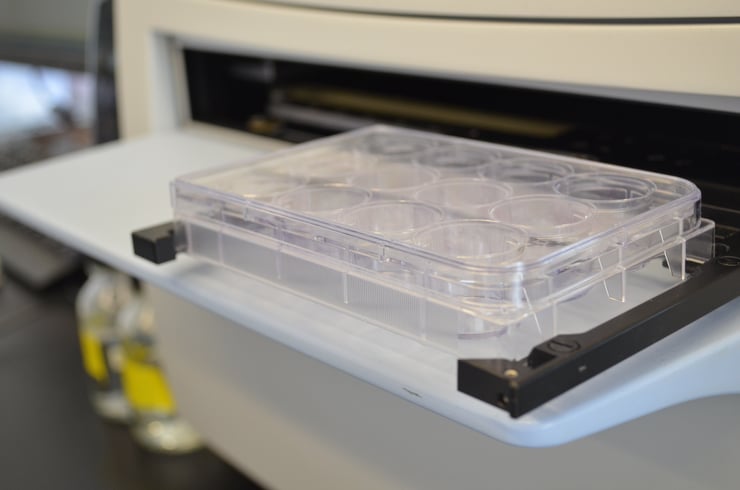
- 24h after transfection, cells are detached using trypsin-EDTA or Versene (for adherent cell layer). Cells are then microcentrifuged 5min at 300g at room temperature. The supernatant is discarded and the cells washed and resuspended in HEPE-buffered DMEM without phenol red. 40-100uL of cells are distributed per well of a 96-well white cell culture plate and maintained at 37 degree (C) with 5% CO2 in a humidified chamber for additional 24h to allow attachment before BRET measurement.
- Detach the cells at 48h after transfections and microcentrifuge for 5min at 300g at room temperature. The cell pellets are washed once with 1mL PBS containing 0.5mM MgCl2, and resuspended in 1mL of the same solution plus 0.1% glucose. Equal amounts of cell suspension are distributed to the 96-well white cell culture plate and proceeded to BRET measurement.
5uM of final coelenterazin concentration is used to initiate the energy transfer. BRET signals are measured immediately after substrate addition using a BRET reader capable of measuring light emitted at the donor and acceptor wavelengths in a quasi-simultaneous manner. As the BRET signal is time-sensitive, only add the substrate up to 12 wells at a time.
For reproducible results, BRET measurements should be performed under temperature-controlled conditions.
Analysis of BRET Signal
Non-specific BRET signals will increase linearly with increasing acceptor concentrations, whereas specific BRET signals will increase in a hyperbolic manner due to the eventual saturation of all donors with acceptors.
As a result, the BRET signal measured has to be corrected for the background signal due to the overlap of donor emission at the acceptor wavelength, which is determined in parallel for cell expressing the donor alone.
BRET Signal = [ Acceptor Emission / Donor Emission ] - cf
where cf, the correction factor, corresponds to [Acceptor Emission / Donor Emission ] from cells transfected with protein-donor alone.
For detailed protocols on how to perform a BRET saturation assay and competition assay, please refer to Borroto-Escuela et al. (2014).
In the next article, we'll continue our discussion on techniques for studying protein-interactions and introduce a technique very similar to BRET but does not require modification of the interacting proteins: Fluorescence Resonance Energy Transfer (FRET).